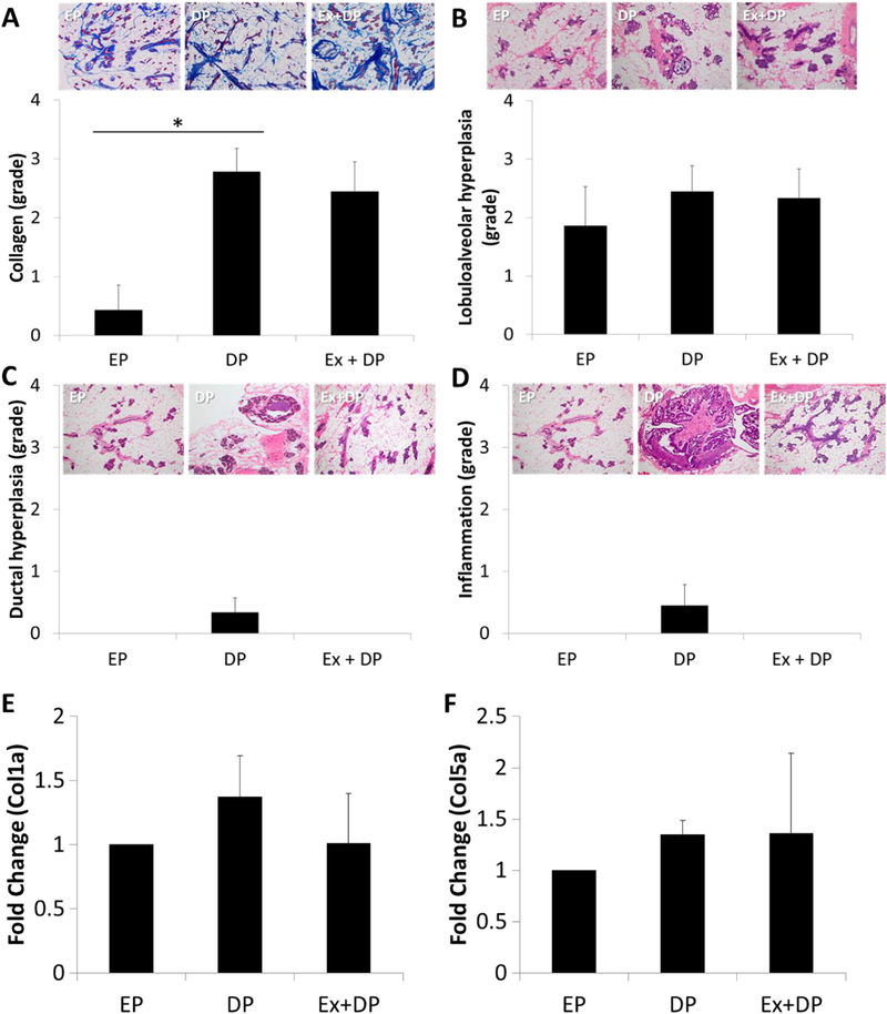
Fig. 2.

Histological assessment of non-tumor mammary glands. Collagen levels (A) (Masson’s trichrome stain - 4×) were significantly decreased in rats with early parity (EP) compared to delayed parity (DP), independent of exercise (Ex). There were no significant differences in lobuloalveolar hyperplasia (B), ductal hyperplasia (C), or inflammation (D). No differences were observed for gene expression levels of collagen 1a (Col1a1) (E) or 5a (Col5a1) (F). H&E stained images are shot at 10×. *p < 0.01.
