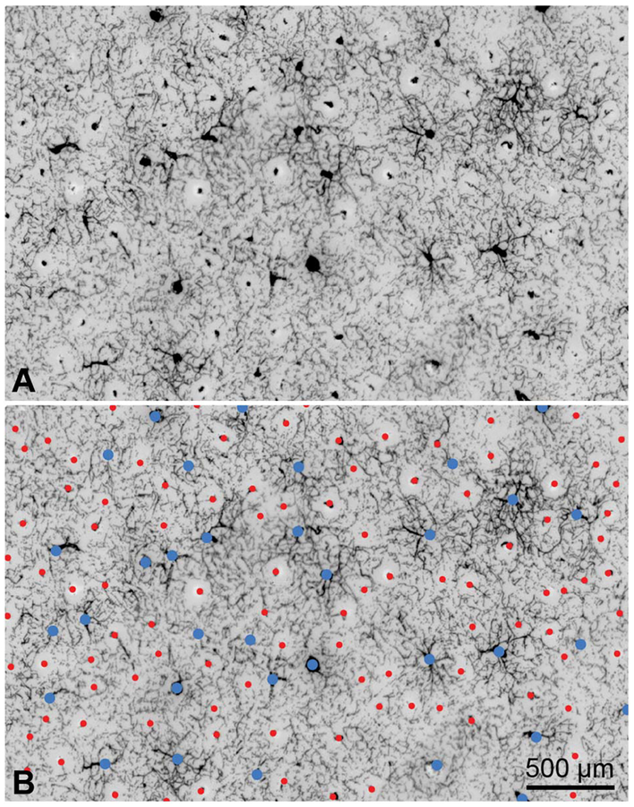
FIG. 1.
Distribution of arterioles and venules in macaque striate cortex. A. A 150-μm tangential section in layer 3 shows profiles of arterioles and venules in cross-section. They are labeled by diaminobenzidine, reflecting the distribution of red blood cells in the unperfused tissue. Arterioles are small and surrounded by pale, capillary-free zones. The venules are larger, with radiating branches that drain the surrounding capillary bed. B. Same image, identifying arterioles (red dots, n = 101) and venules (blue dots, n = 34). Each microvascular lobule comprises an irregular array of arterioles surrounding a single venule or, occasionally, pair of venules.

