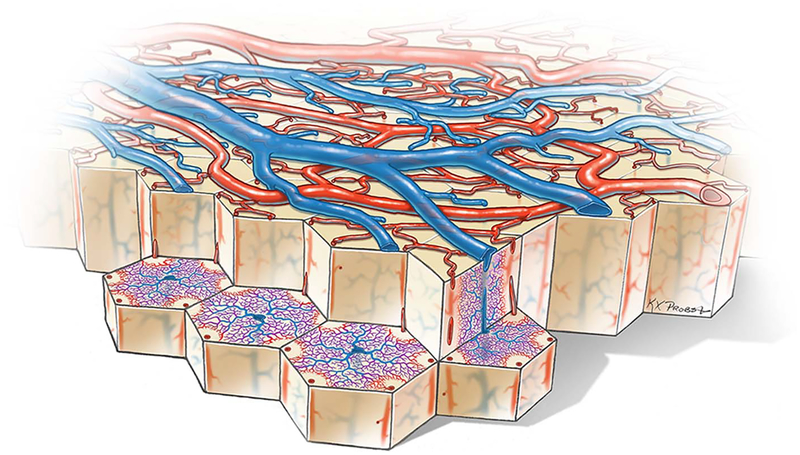
FIG. 3.
Cutaway diagram reveals vascular microlobules of the cerebral cortex, each comprised of an approximately hexagonal array of penetrating arterioles that supply the capillary bed, which drains into a central venule. A cylindrical capillary-free zone surrounds each penetrating arteriole. The surface arterioles form a reticulum to allow efficient delivery of blood to satisfy local fluctuations in metabolic demand. Flow is controlled by a system of sphincters located at both arteriolar junctions and penetration sites of descending arterioles. The anastomotic organization of the surface arterioles renders the cortex resistant to microinfarction by emboli because after occlusion of a vessel, any given location can still be perfused by blood flowing from another direction. Even occlusion of a penetrating arteriole may be tolerated by collateral flow through the capillary bed.

