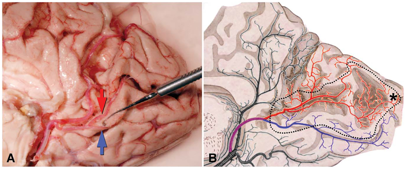
FIG. 4.
Arterial circulation of the primary visual cortex. A. Medial view of the right occipital lobe with the inferior lip of the calcarine sulcus deflected downward by a muscle hook to show the bifurcation of the calcarine artery. The superior calcarine artery(red arrow) crosses the calcarine sulcus to feed the upper calcarine bank and a portion of the cuneus. The inferior calcarine artery (blue arrow) supplies the lower calcarine bank and some of lingual gyrus. B. Drawing of the occipital circulation, after Polyak (40), demonstrates territories supplied by the superior (red) and inferior (blue) calcarine arteries. He incorrectly shows the arteries bifurcating serially until they end in terminal branches. Dotted line corresponds to perimeter of primary visual cortex. *Foveal representation.

