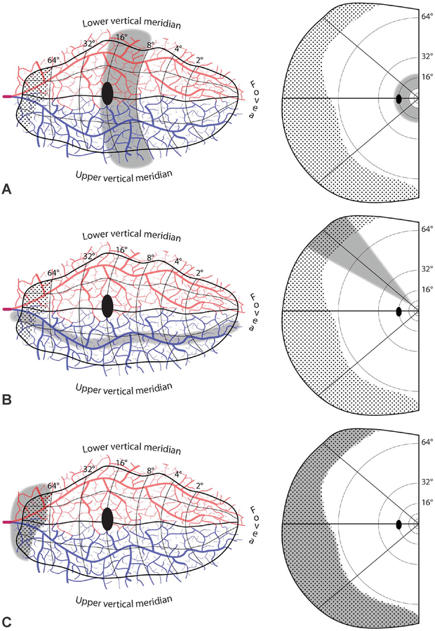
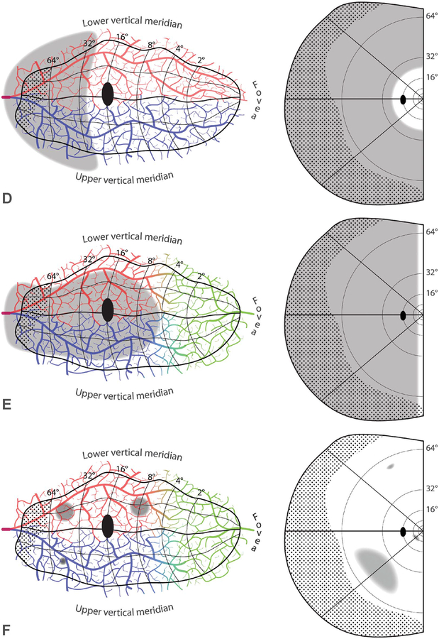
FIG. 6.
Disallowed embolic cortical visual field defects. A. Hemiannular scotoma, from an infarct that crosses vascular territories. B. Sectoranopia from a long, thin infarct running roughly parallel to an isopolar ray in the visual field map. C. Missing temporal monocular crescent, from infarct confined to its representation (dotted region). D. Peripheral scotoma, sparing the central 24°.E. Homonymous hemianopia, sparing a vertical strip of uniform azimuth along the vertical meridian. F. Isolated homonymous scotomata, from focal cortical infarcts. In these schematic examples, two ~5-mm infarcts are shown from an embolus imagined to arrest surface arteriolar flow despite availability of collaterals. They would produce scotomas in the inferior visual field of vastly different size. A microinfarct from occlusion of a penetrating vessel is also shown, with a corresponding microscotoma in the peripheral upper field. Visual fields riddled with homonymous microscotomata from emboli are not encountered in clinical practice.


