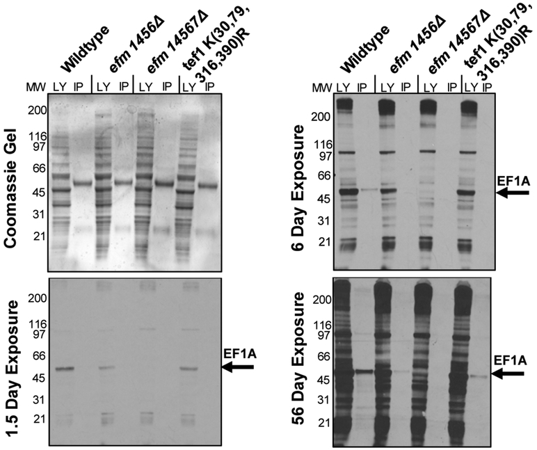
Figure 2: Immunoprecipitation of EF1A from methylation-deficient cells shows specificity of elongation factor methyltransferases.
Yeast cells from wildtype and mutant strains that were labeled with S-adenosyl-[methyl-3H]methionine, and immunoprecipitated with an anti-EF1A polyclonal antibody as described in “Experimental Procedures”. The top left panel is a Coomassie-stained polyacrylamide gel, which serves as a protein loading control. The remaining panels show the detection of radioactive material in each sample at different time intervals. The longer exposure reveals that EF1A methylation is decreased in the methyltransferase knockout mutants. The LY lane shows the total lysate before the immunoprecipitation while the IP lanes show what was pulled down with the EF1A-antibody. The figure shown is a representative from one out of two separate experiments.

