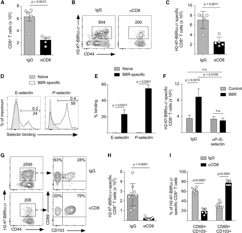
Figure 3. Circulating Memory CD8+ T Cells Are Recruited to the Site of TRM Activation and Are the Source of Secondary Trm CD8+ T Cells.
(A–C) Mice were infected on the left ear skin with VACV and received control IgG or CD8 depleting antibody 35 days post-infection. Mice were then challenged with B8R20–27, and the number of CD8+ T cells (A) and B8R20–27-specific CD8+ T cells (B and C) was quantified 40 h after challenge. (B) Representative flow cytometry plots depicting B8R-specific CD8+ T cells in the challenged skin. (C) Quantification of (B).
(D) Mice were infected on the skin with VACV, and on day 35 after infection, B8R-specific CD8+ T cells in the blood were analyzed for expression of P- and E-selectin ligands. Naive CD8+ T cells (CD62L+/CD44−) were used as a negative control for selectin binding.
(E) Quantification of (D).
(F) Mice were infected on the skin with VACV and received control IgG or P-/E-selectin blocking antibodies before challenge with B8R20–27 or control peptide on day 40 post-infection. The number of B8R-specific CD8+ T cells in the skin 40 h after challenge was determined by flow cytometry (see Figure S3).
(G) Mice were treated as in (A), except they were challenged three times with B8R20–27 (as in Figure 2E) and analyzed by flow cytometry 10 days after the last peptide challenge.
(H) Quantification of the number of B8R-specific CD8+ T cells in (G).
(I) Quantification of CD103 and CD69 expression by B8R-specific CD8+ T cells identified in (G).
Statistical significance was determined using an unpaired t test. Error bars represent SEM. Data in (A)–(C) are representative of 2 independent experiments (n = 3–5). Data in (D)–(F) are representative of 3 independent experiments (n = 3–10). Data in (G)-(I) are pooled from 2 independent experiments (n = 4–5).

