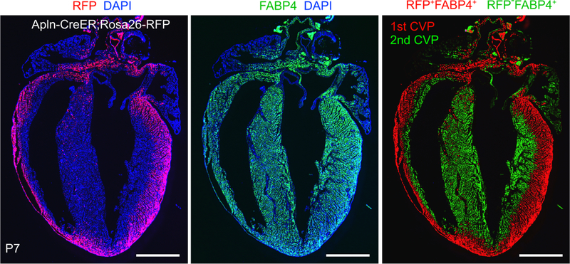
Figure 6. 1st and 2nd coronary vascular population in neonatal heart.
Apln-CreER, induced at E10.5, labeled fetal coronary VECs with Cre-activated RFP expression. In the P7 postnatal heart, VECs were present throughout the myocardial wall, as demonstrated by immunostaining for the VEC-selective marker FABP4 (middle panel), but the RFP lineage tracer of fetal VECs was only observed in the outer myocardial wall (left panel). The FABP4+RFP+ VECs, descended from fetal VECs, are designated the 1st coronary vascular population (CVP; red pseudocolor, right panel) while the remaining FABP4+RFP− VECs, formed de novo in the postnatal heart, are designated the 2nd CVP (green pseudocolor, right panel). Bar = 1 mm.

