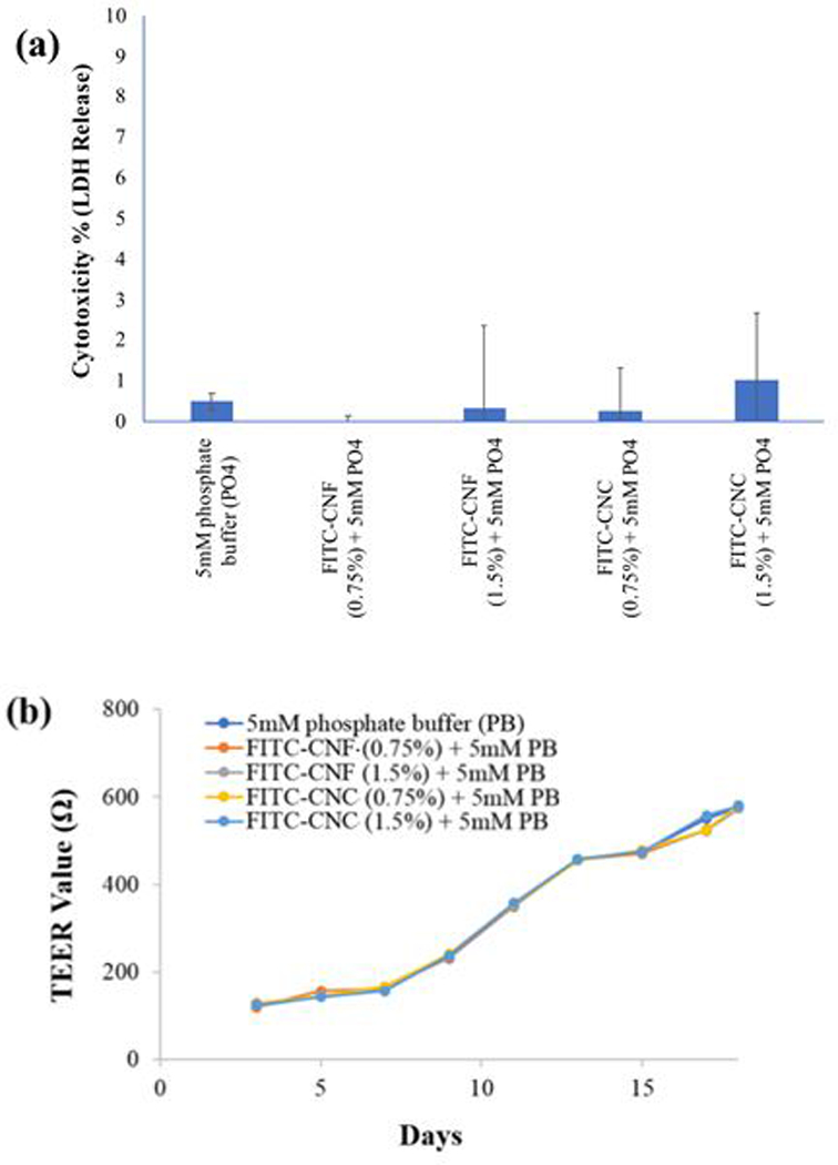
Figure 7.

(a) Lactate dehydrogenase release (cytotoxicity percentage): LDH release analysis showed no membrane damage to the GIT tri-culture cells following 24 h exposure to the FITC-CNF and FITC-CNC digesta. (b) Transepithelial electrical resistivity (TEER) analysis: a gradual increase in TEER values indicated that the cell junctions of the cellular monolayer remained intact during 17 days of cell culture. On day 18, cells were exposed for 24 hrs to the FITC-CNF and FITC-CNC digesta and the TEER values across the transwell membranes did not decrease, thus indicating no loss of cell viability or loss of cellular monolayer integrity had occurred.
