Figure 1.
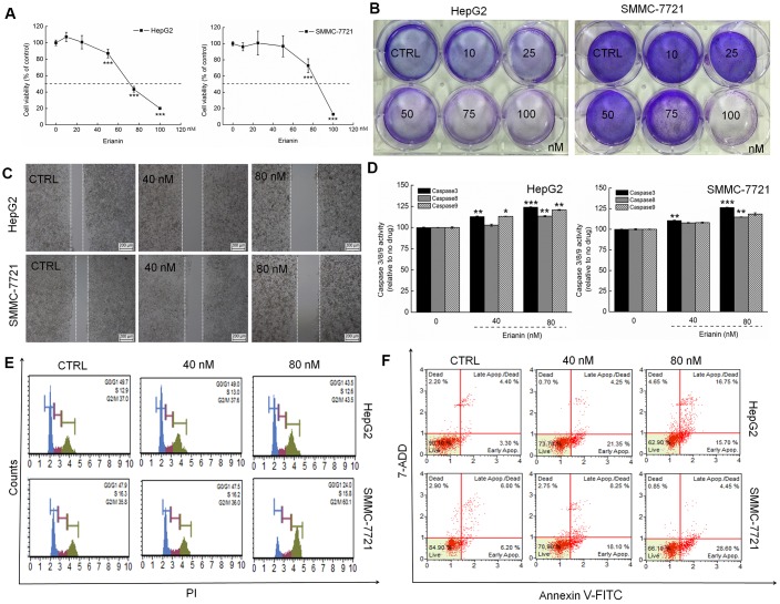
Erianin showed toxicity toward liver cancer cells. (A) Erianin reduced HepG2 and SMMC-7721 cell viability in a dose-dependent manner after a 24-h treatment. (B) Erianin significantly inhibited the formation of HepG2 and SMMC-7721 cell colonies (crystal violet staining, n = 6). (C) Erianin inhibited HepG2 and SMMC-7721 cell migration (migration assay, n = 6; 4× magnification, scale bar: 200 μm). (D) Erianin enhanced caspase-3, -8, and -9 activation in HepG2 and SMMC-7721 cells. Data are expressed as percentages relative to the corresponding control cells and as mean ± SD (n = 6). *P < 0.05, **P < 0.01, and ***P < 0.001 vs control cells. (E) Erianin increased the G2/M phase proportion within the cell cycle distribution (n = 6). (F) Erianin induced liver cancer cell apoptosis (n = 6).

