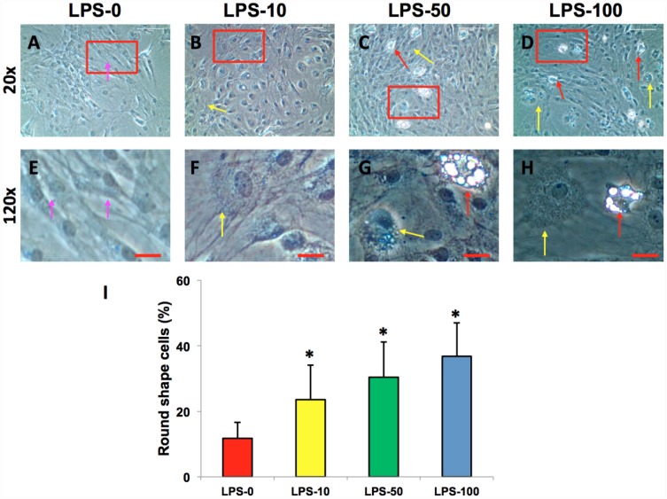
Figure 3.
LPS effect on the proliferation and differentiation of rabbit AF cells cultured for 7 days. LPS not only decreased the proliferation of rabbit AF cells, but also induced significantly morphological change in rabbit AF cells during the culture (A–H). The results showed that the cell number was decreased with increasing the concentration of LPS. Many cells have changed their shape from spindle shape to round shape (yellow arrows in B–D, F–H). Some adipocyte-like cells were also found in the LPS-treated groups (red arrows in C, D, G, H). Semi-quantification indicated that the round shape cells were increased at a LPS concentration dependent manner (I). The images of E–H were the enlarged images of the box areas in the images of A–D. *p<0.05 compared to the control group. White bars = 200 μm, red bars = 50 μm.

