Figure 4.

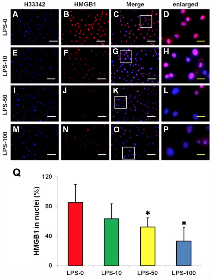
LPS effect on HMGB1 releasing from the nuclei to the cytoplasma of the rabbit AF cells cultured for 7 days. (A–D) normal rabbit AF cells (LPS-0). (E–H) the rabbit AF cells were treated with 10 ng of LPS (LPS-10). (I–L) the rabbit AF cells were treated with 50 ng of LPS (LPS-50). (M–P) the rabbit AF cells were treated with 100 ng of LPS (LPS-100). (Q) semi-quantification of positive stained HMGB1 in the nuclei of the cells. More than 95% of the nuclei of the normal cells were positively stained with HMGB1 (B–D, Q). LPS induced HMGB 1 releasing from the nucleus of rabbit AF cells to the cytoplasma (F–H, J–L, N–P). Semi-quantification indicated that the concentration of HMGB1 in the cytoplasma increased with increasing of the concentration of LPS (Q). (A, E, I, M) H33342 staining. (B, F, J, N) anti-HMGB1 antibody staining*p<0.05 compared to the control group. (C, G, K, O) merged images of H33342 stained images and anti-HMGB1 antibody stained images. (D, H, L, P) enlarged images of the box areas in the images of (C, G, K, O). White bars = 100 μm, yellow bars = 25 μm.
