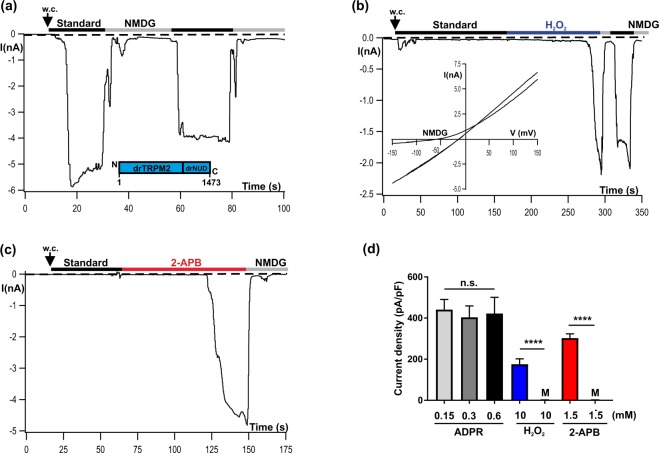
Figure 1.
Activation of drTRPM2 by ADPR, H2O2 and 2-APB. Representative whole-cell patch-clamp experiments of HEK-293 cells heterologeously expressing drTRPM2 (a) ADPR (0.15 mM) was infused into the cell through the patch pipette together with 1 µM Ca2+. Current onset occurs with a short delay after reaching whole-cell configuration (w.c.). Substitution of external Na+ in the standard bath solution (indicated with black bars, ref. to Methods) with the impermeable cation NMDG (indicated with gray bars) blocks the inward currents. (b) Same pipette solution as used in panel a but without ADPR. Activation is induced after extracellular application of 10 mM H2O2 to the standard bath solution (indicated with blue bar). Note the delayed time course of activation. The corresponding current-voltage relation, as obtained with voltage-ramps, is given in the inset. (c) Same pipette solution as used in panel b. Stimulation was performed by superfusion of the cells with standard bath solution containing 1.5 mM 2-APB (indicated with red bar). There was no noticeable current decline unless the cells were superfused with NMDG bath solution. (d) Summary of the effects of different agonists on drTRPM2 including control experiments (M, Mock-transfected cells) either performed with ADPR or with 2-APB. All data are presented as mean ± s.e.m. Differences are significant at ****(P < 0.0001) evaluated with an unpaired Student’s t-test, n = 4–16. n.s., not significant.

