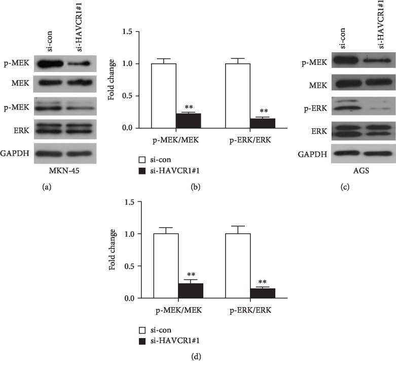
Figure 5.
Activation of MEK/ERK signaling pathway was associated with HAVCR1 expression. (a) The MEK/ERK related protein levels were examined by western blot analysis in MKN-45 cells after si-HAVCR1#1 transfection. (b) The gray values of protein bands were quantified. ∗∗p < 0.01 versus the si-con group. (c) The effects of HAVCR1 deficiency in MEK, p-MEK, ERK, and p-ERK expression levels in AGS cells were measured by western blot. (d) The expression levels of the above proteins were quantified and normalized to GAPDH. One specific siRNA (si-HAVCR1#1) was used as an experimental group because of its higher knockdown efficiency, and nonspecific siRNA was used as a negative control group (si-con). ∗∗p < 0.01 versus the si-con group.

