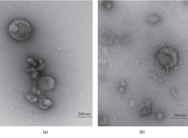
Figure 4.
Transmission electron microscopy displaying exosomal morphology and approximate size. Representative electron micrographs of pooled exosome isolations by size exclusion chromatography. (a, b) The presence of intact spherical/donut-shaped particles, for both bovine and human exosomes, respectively. Scale bar 200 nm. (a) Bovine. (b) Human.

