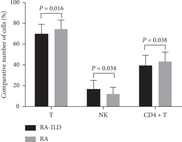
Figure 2.

Relative counts of CD3−CD56+ NK, T cells, and CD4+ T cells between RA-ILD and RA patients. Comparing the peripheral lymphocyte subsets between RA-ILD and RA patients, the percentage of CD3−CD56+ NK cells in the RA-ILD group is higher than that in the RA group. The number of T cells and CD4+ T cells in RA-ILD group is lower than that in the RA group. The data are shown as the means, and the error bars represent the SD. Shown are the significant differences assessed by the binary logistic regression model.
