Figure 7.
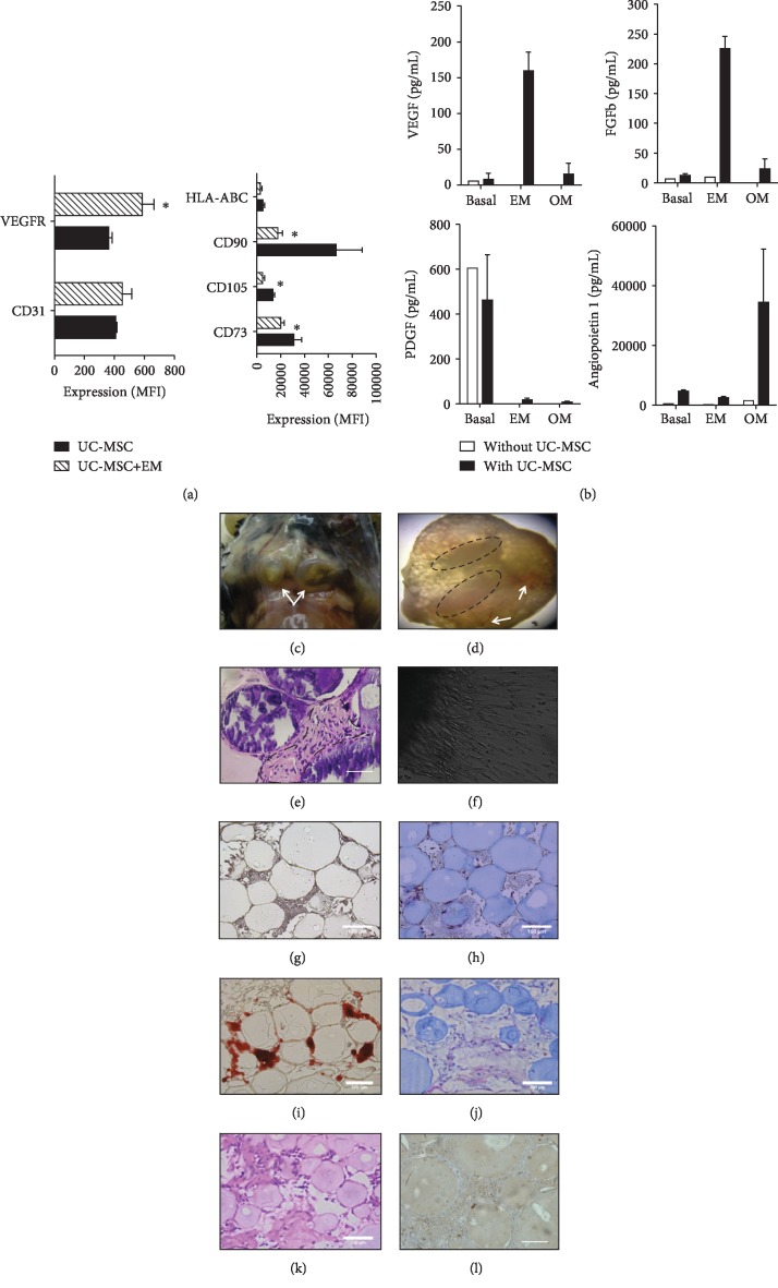
In vivo formation of bone-like scaffolds based on UC-MSC. (a) Expression of endothelial and MSC markers in cells treated with endothelial induction (EM) or growth media. (b) Concentration of growth and angiogenic factors in EM, OM-treated UC-MSC, or nontreated (basal) controls. (c) Bone-like tissue formation at 12 weeks after transplantation of UC-MSC-microbeads in mice. (d) Mineralization zones are evident at the upper part (circles) and angiogenesis (arrows) in bone-like tissue formed from UC-MSC. (e) Hematoxylin and eosin staining evidence the presence of osteoid cells of immature appearance (dotted line) in bone-like tissue formed from UC-MSC scaffolds. (f) Cell migration outside explants of in vivo-formed bone-like tissue after 48 h of culture. Representative micrographs of bone constructs in growth medium (g) and differentiation medium (h) stained for Alizarin Red after 14 days of culture. Representative micrographs of bone constructs without (i) and with UC-MSC (j) stained with Masson's trichrome cultured for 14 days. Micrographs of hematoxylin and eosin (k) and collagen (l) staining of 14-day cultured scaffolds containing UC-MSC. Differences between expressions of different markers were compared. ∗ indicates p < 0.05. Bars indicate 100 μm.

