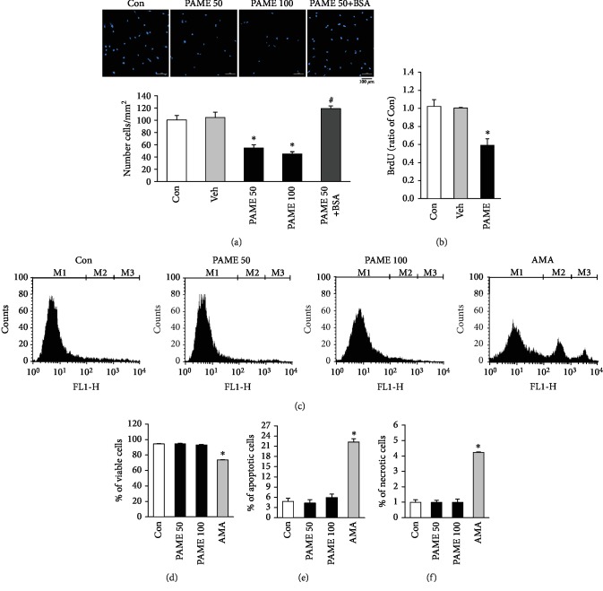
Figure 2.
Effects of PAME on cell growth and death in hBM-MSCs. (a) Treatment with PAME (50 and 100 μM) for 48 h inhibited hBM-MSC proliferation. This inhibition was blocked by 0.5% BSA (n = 3). (b) PAME (50 μM)-induced proliferation inhibition in hBM-MSCs was confirmed by BrdU assay (n = 7). (c) Histogram plots of viable (M1), apoptotic (M2), and necrotic cells (M3) distinguished by flow cytometric analysis using Annexin V-FITC and SYTOX green dye staining. The (d) viable cells, (e) apoptotic cells, and (f) necrotic cells were not significantly different between the PAME-treated group and the control group (n = 6). AMA served as a positive control for cell death induction (n = 3). All data represent mean ± SEM. ∗p < 0.05, versus the control group; #p < 0.05, versus the PAME group. Con: control; AMA: antimycin A; BSA: bovine serum albumin; Veh: vehicle.

