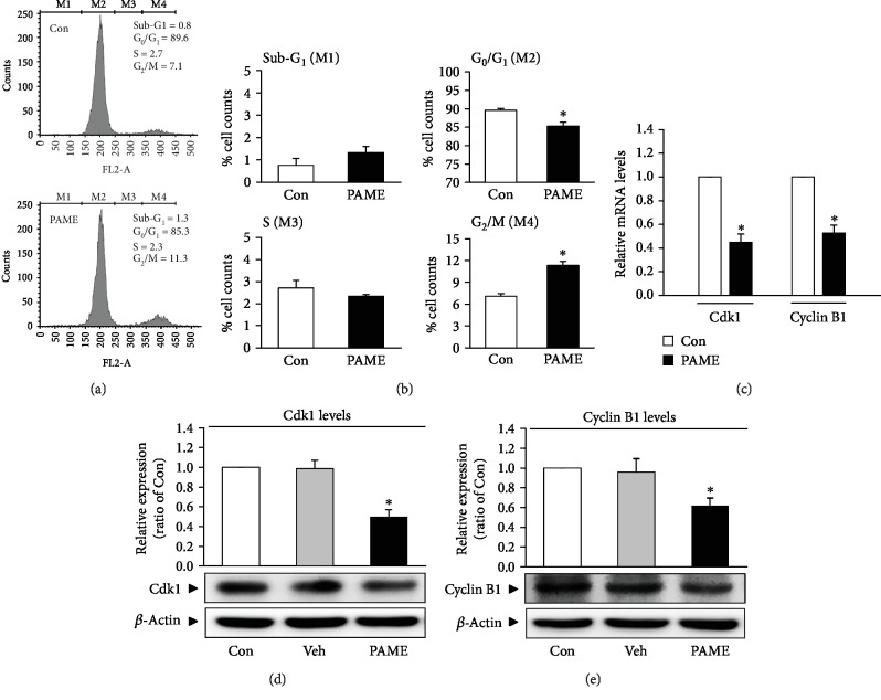
Figure 3.
PAME induces G2/M cell cycle arrest in hBM-MSCs. The cells were treated with PAME (50 μM) for 48 h prior to the flow cytometric and Western blot analyses. (a) Representative histograms of cell distribution at the sub-G1 phase (M1), G0/G1 phase (M2), S phase (M3), and G2/M phase (M4) detected by flow cytometry using PI staining. (b) PAME (50 μM) significantly increased the cell population at the G2/M phase (M4) and decreased at the G0/G1 phase (M2) (n = 8). PAME significantly decreased the levels of (c) Cdk1 and cyclin B1 mRNA (n = 8) and (d, e) protein (n = 5). The levels of mRNA and protein were examined by qRT-PCR and Western blot analyses, respectively. All data represent mean ± SEM. ∗p < 0.05, versus the control group. Con: control; Veh: vehicle; PI: propidium iodide.

