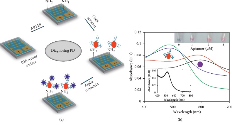
Figure 1.
(a) Schematic representation of alpha-synuclein detection by an aptamer-gold nanoparticle probe on an IDE surface. The surface was modified by APTES to immobilize the GNP-aptamer, and then, alpha-synuclein was detected. (b) Colorimetric analysis to reveal the stability of aptamer attachment on the GNPs. Analyses were performed by both colorimetric (figure inset) and spectrophotometric methods. The figure inset graph represents the absorbance maximum of as-received GNPs.

