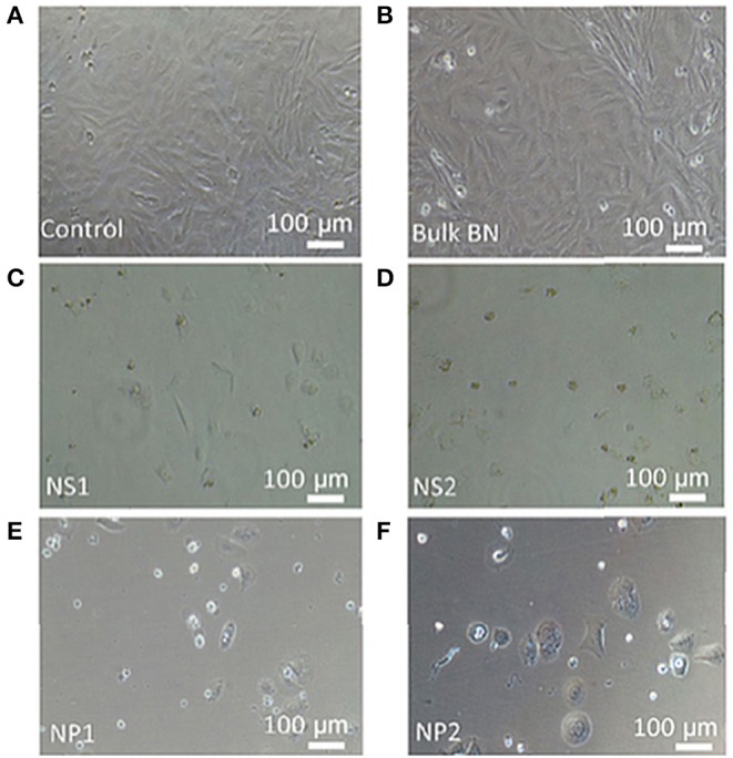
Figure 4.

Bright-field microscopy images of SaOS2 cells cultured in the presence of (A) standard culture medium (control), (B) bulk BN, (C) nanosheet NS1, (D) nanosheet NS2, (E) nanoparticle NP1, and (F) nanoparticle NP2. Reproduced with Permission from Mateti et al. (2018).
