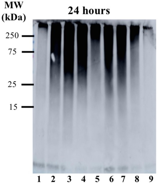
Figure 5.

Gel electrophoresis/Western blot of 25 μM Aβ1−42 and different concentrations of Ru-N-1 and Ru-N-4 in PBS buffer (0.01 M, pH 7.4) at 24 h incubation with agitation at 37°C, using anti-Aβ antibody 6E10. Lane 1: Aβ1−42; lane 2: Aβ1−42 + 0.25 eq. Ru-N-1; lane 3: Aβ1−42 + 0.5 eq. Ru-N-1; lane 4: Aβ1−42 + 1 eq. Ru-N-1; lane 5: Aβ1−42 + 2 eq. Ru-N-1; lane 6: Aβ1−42 + 0.25 eq. Ru-N-4; lane 7: Aβ1−42 + 0.5 eq. Ru-N-4; lane 8: Aβ1−42 + 1 eq. Ru-N-4; lane 9: Aβ1−42 + 2 eq. Ru-N-4.
