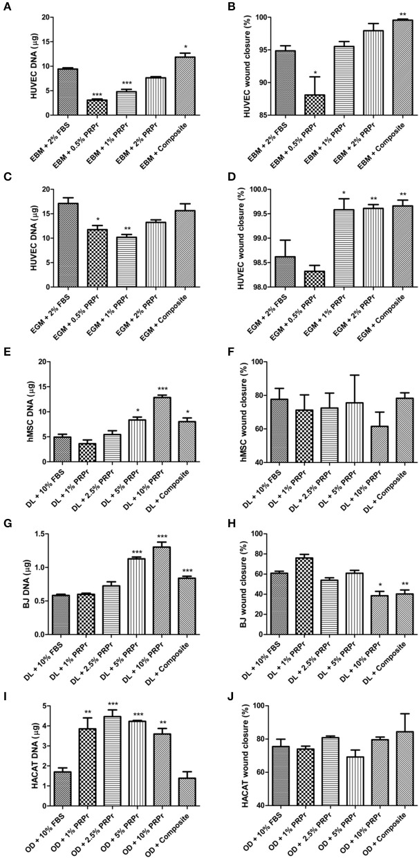
Figure 4.
Platelet-rich plasma releasate (PRPr) and composite scaffolds sustain cell proliferation and migration. Cells were cultured in control medium supplemented with FBS or different concentrations of PRPr. Additionally, cells were cultured in FBS-free medium but with composite scaffold placed in hanging inserts. Proliferation analysis was measured by DNA quantification with picogreen assay after 4 days of culture (A,C,E,G,I) and cell migration was measured by percentage of wound closure in a scratch assay (B,D,F,H,J). HUVEC were cultured in endothelial basal medium (EBM, which sustains cells) (A,B) or endothelial growth medium (EGM, which has a cocktail of factors to promote cell activity) (C,D). hMSC (E,F), and BJ fibroblasts (G,H), were cultured in DMEM low glucose (DL). HACAT were cultured in Optimized DMEM (OD) (I,J). Data are expressed as mean ± standard error of the mean. *p < 0.05, **p < 0.01, and ***p < 0.001 after one-way Anova with Dunnett's post-test analysis.

