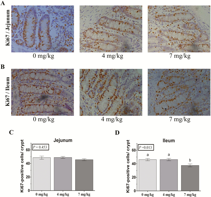
Figure 1.
Post-B6 supplementation weaned piglet intestinal cell proliferation. (A and B) The representative images of immunohistochemistry (IHC) staining with Ki67 antibody in weaned piglet jejunum and ileum shown (×200; n = 6). (C and D) The statistical analysis of Ki67-positive cells in each crypt from images shown on the (A and B). The data were analyzed by one-way ANOVA using the GLM procedure of SAS. a,bWithin a variable, values with different superscripts differ (P < 0.05). Data are expressed as means ± SEM; SEM = standard error of the mean. n = 6.

