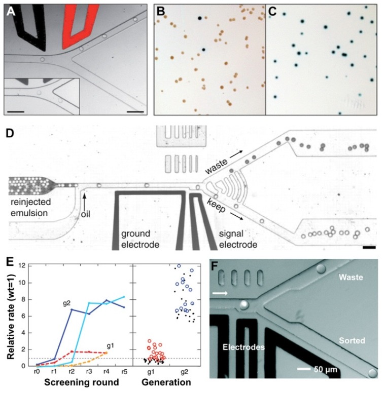
Figure 4.
Fluorescence-activated droplet sorting systems. (A) Trajectories of droplets flowing through the sorting junction of a FADS system. When an AC electric field is applied across the electrodes, the droplets are deflected into the ‘positive’ arm. In the absence of the electric field, the droplets flowed into the ‘negative’ arm by default (inset). (B,C) Enrichment of cells by FADS based on β-galactosidase activity. Photographs of E. coli colonies before (B) and after sorting (C). The lacZ bacteria (blue colonies) were completely purified from the ΔlacZ bacteria (white colonies) after sorting, resulting in only lacZ colonies growing on the agar. Figure 3A–C were adapted from Reference [30] with permission from the Royal Society of Chemistry. (D) A different design of the FADS system. The droplets flow as a solid plug to a junction where oil is added to space the droplets. Light droplets contain 1 mM fluorescein and the dark ones contain 1% bromophenol blue. When a droplet passes by the laser that is focused on the channel at the gap between two electrodes, its fluorescence intensity is detected. If the intensity is above a threshold (in the case of light droplets), the droplet is sorted by dielectrophoresis towards the bottom channel. (E) Enrichment of library pools. The activities are normalized relative to wild-type HRP. The first-generation epPCR and saturation mutagenesis libraries (dashed red and orange, respectively) enrich to a level of about two times the activity of the wild type after four sorting rounds. The second-generation low- and high-mutation rate libraries (solid blue and cyan, respectively) enrich to about eight times the wild type. The right panel shows a dot plot of the activities of the 50 unique first-generation (g1) mutants (red circles) and 31 second-generation (g2) mutants (blue circles). Figure 3D,E were adapted from reference [19], complying with the License for PNAS Articles. (F) The sorting junction of another FADS system. Single droplet fluorescence was detected following excitation by the laser (the white dot). The default flow path of the droplet is towards the top ‘waste’ channel since the waste outlet is at atmospheric pressure and a withdrawal of less than half the total flow rate is applied to the ‘sorted’ outlet. However, if the droplet fluorescence exceeds a predefined threshold an electric field is activated between the electrodes, pulling the droplet to the bottom channel. Figure 3F was adapted from Reference [47] with permission from the Royal Society of Chemistry.

