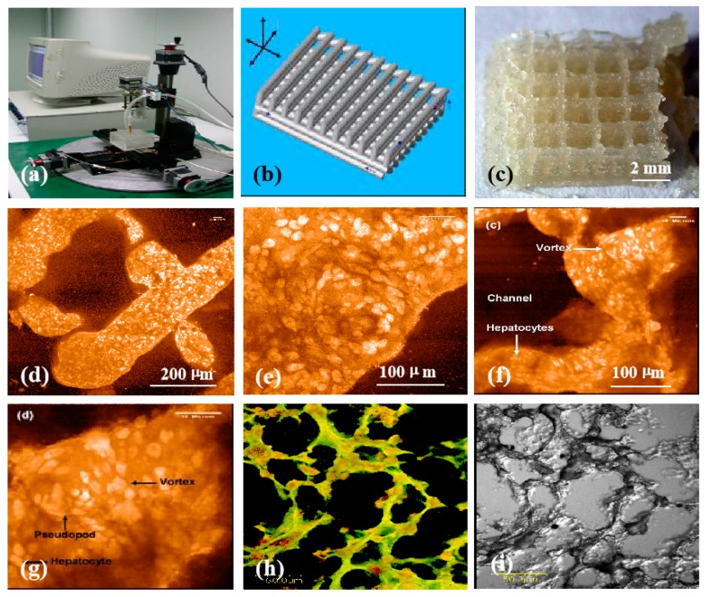
Figure 9.
Three-dimensional (3D) printing of hepatocyte-laden chitosan/gelatin hydrogels and laser scanning confocal microscope observations of hepatocytes in the 3D printed chitosan/gelatin constructs with propidium iodide staining: (a) The 3D bioprinter made in the corresponding author’s laboratory in 2003; (b) a grid computer-aided design (CAD) model; (c) a grid cell-laden 3D construct immediately after 3D bioprinting; (d,e) hepatocytes in the 3D printed chitosan/gelatin construct 1 month after in vitro culture; (f–i) hepatocytes in the 3D printed chitosan/gelatin construct 2 months after in vitro culture; (e,g) are the magnifications of (d) and (f) respectively; (i) a dark-field micrograph of (h). Image reproduced with permission from [154].

