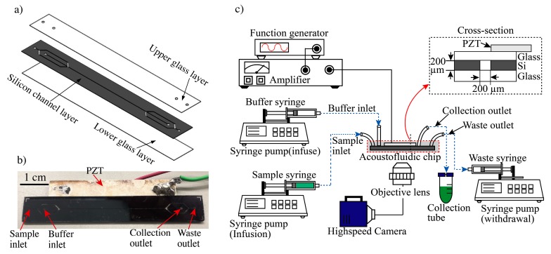
Figure 3.
Experimental setup of the acoustofluidic system for cell separation and collection. (a) Structure of acoustophoresis microdevice. (b) Real picture of the device. (c) A sample syringe pump and buffer syringe pump infuse sample or buffer solution into the inlet ports. A separate syringe pump withdraws fluid designated as waste. Separated target samples are collected through the collection tube for downstream analysis. A function generator with a power amplifier applies the AC power to the piezoelectric actuator to generate an acoustic radiation force in the microchannel. This force allows for particle focusing and separation of samples. A high-speed camera module allows for visualization of the separation.

