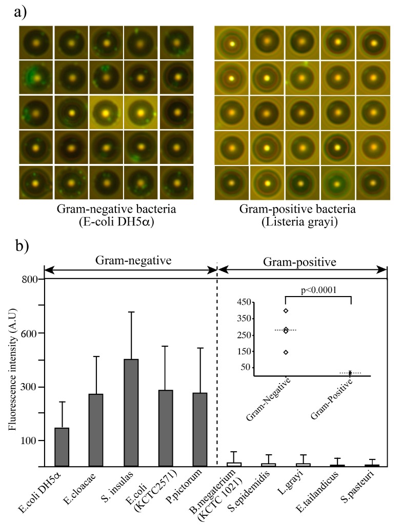
Figure 5.
Binding profile of aptamer-modified microbeads against gram-negative and gram-positive bacteria. (a) Collected beads from collection outlet. Gram-negative bacteria are bound on the aptamer-coated bead while Gram-positive bacteria shows no or less binding on the beads. Fluorescent intensities were measured each bead and summarized the intensities. After fluidic operation, the target sample and waste were collected into tubes and the waste outlet syringe, respectively. Then, 10-µL samples were taken from the sample collection tubes and dropped onto slide glasses for observation of fluorescence intensity using a fluorescence microscope. (b) The fluorescence intensity of the beads (>100 beads) was measured using Image J software (NIH). Data are shown as means ± SD of three independent experiments. Signal intensity was significantly different between gram-negative and gram-positive bacteria (p < 0.0001, t-test).

