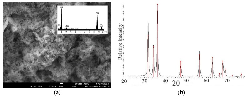
Figure 4.
(a) Scanning electron microscopy (SEM) shows zinc oxide nanoparticles forms (ZnO NPs) foliarly applied to foxtail millet. Inset: The electron dispersive analysis (EDAX) profile where Zn and O are detected as the major elements. (b) X-ray diffraction analysis verified that the zinc oxide nanoparticles have wurtzite-structural symmetry.

