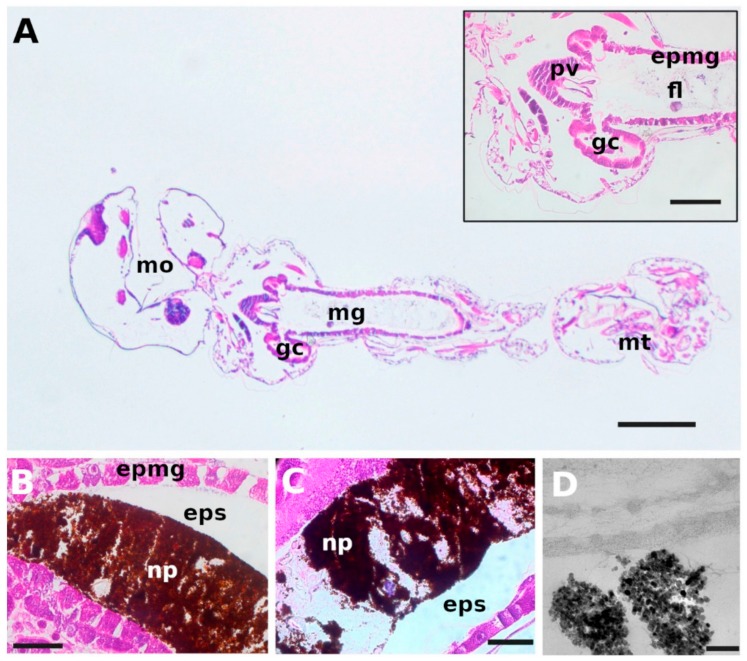
Figure 8.
Microscopic characterization of Aedes aegypti larvae. (A) Optical micrograph of the sagittal section of a control sample. Inset: Optical micrograph of the section of gastric caeca, proventriculus and anterior end of mid-gut. (B) Optical micrograph of the midgut of a larva after exposure to 100 mg L−1 BIONs for 1 h, evidencing the accumulation of nanoparticles in the gut lumen. (C) Optical micrograph of the midgut of a larva exposed to 100 mg L−1 BION@chlorin for 80 min. (D) TEM micrograph of the gut of a Aedes aegypti larva exposed to 100 mg L−1 BION@chlorin for 1 h. (epmg), epithelium of the midgut; (eps), ectoperitrophic space; (fl), food in gut lumen; (gc), gastric caeca; (mg), midgut; (mo), mouth; (mt), malpighian tubules; (pv), proventriculus; (np), nanoparticles. Size bars: A = 250 μm, inset = 100 μm, B and C = 50 μm, D = 200 nm. Reproduced from [99], with permission from Elsevier, 12019.

