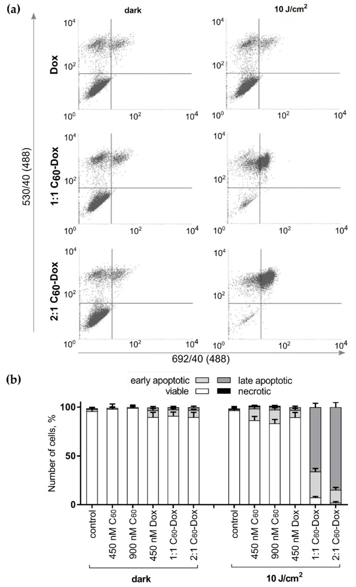
Figure 8.
Cell death differentiation in CCRF-CEM treated with either free C60, Dox or C60-Dox nanocomplexes: (a) flow cytometry histograms of CCRF-CEM cells stained with Annexin V-FITC/ propidium iodide (PI) after treatment either with C60-Dox alone or in combination with 405 nm light (in each panel the lower left quadrant shows the content of viable, upper left quadrant—early apoptotic, upper right quadrant—late apoptotic, lower right quadrant—necrotic cells populations); (b) Quantitative analysis of cell population content, differentiated with double Annexin V-FITC/PI staining.

