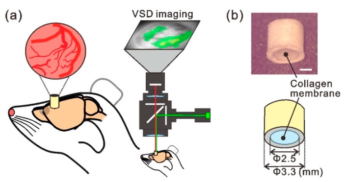
Figure 1.
Experimental concept of the implantable cranial window for chronic voltage-sensitive dye (VSD) imaging. (a) Schematic view of the experimental setup. A cylinder-like device was attached to the skull and conventional VSD imaging [21] was performed by peering through it. (b) Upper image: photograph of the tube-shaped implantable-chamber device, wherein the bottom end of the tube was sealed with the atelocollagen membrane. The device was printed using a high-resolution three-dimensional printer. Lower image: design drawing of the device. Scale bar in (b), 1 mm.

