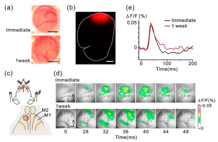
Figure 3.
Forelimb-evoked VSD responses recorded through the implanted-cranial window. (a) Representative images of the brain surface in different rats obtained immediately (upper) and 1 week (lower) after the implant surgery. (b) Histological fluorescent assessment of VSD diffusion into the cortex through the collagen membrane. Red areas indicate the cortex stained by the VSD-RH795. Dotted line indicates the contour of the brain section. (c) Schematic view of the experiments. The implanted device was placed over M2. (d) Forelimb-evoked neuronal responses were recorded immediately (upper panels) and 1 week (lower panels) after implanting the chamber device. Data are from different animals. (e) Representative time course of the optical signal evoked by forelimb stimulation. Scale bars in (a,b,d), 1 mm. M1, primary motor cortex; M2, secondary motor cortex; A, anterior; L, lateral.

