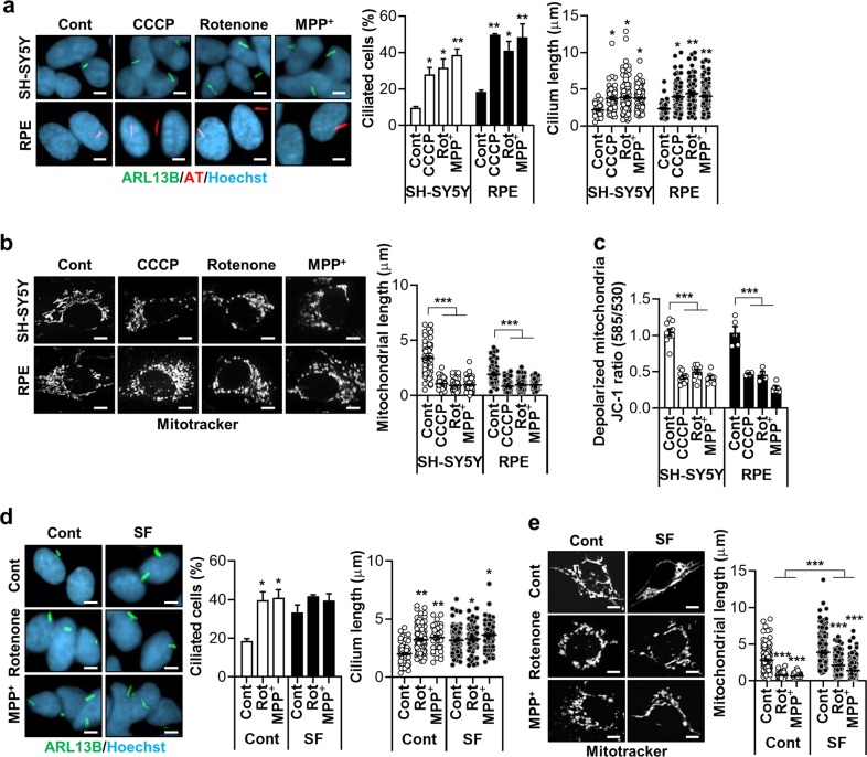
Fig. 1. Mitochondrial respiratory inhibitors stimulate ciliogenesis.
a Increased ciliogenesis occurs following treatment with the mitochondrial respiratory inhibitors CCCP (5 μM), rotenone (200 nM), and MPP+ (5 mM) in (upper) SH-SY5Y and (lower) RPE cells. Cells were cultured to almost 100% confluency and treated with these agents for 24 h. Primary cilia were stained with antibodies against ARL13B (green) or acetylated α-tubulin (AT) (red) and the nucleus (blue) was counterstained with Hoechst 33342 dye. b CCCP (5 μM), rotenone (200 nM), and MPP+ (5 mM) were applied to (upper) SH-SY5Y and (lower) RPE cells and mitochondria were stained with a MitoTracker (white) to measure mitochondrial length. c SH-SY5Y and RPE cells were treated with CCCP (5 μM), rotenone (200 nM), and MPP+ (5 mM). After 24 h, the alteration of mitochondrial membrane potential was measured with the MitoProbe JC-1 assay using the Attune NxT flow cytometer. d, e SH-SY5Y cells were treated with rotenone or MPP+ in the presence or absence of serum [normal (Cont) or serum-free (SF)]. Afterward, the ciliated cells and mitochondrial length were determined. Data are the mean ± SEM. *p < 0.05, **p < 0.01, ***p < 0.005 vs. untreated controls determined by ANOVA followed by a post hoc LSD test. Scale bar, 5 μm.

