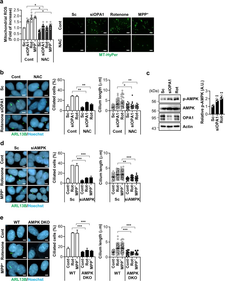
Fig. 3. Mitochondrial ROS and AMPK mediate mitochondrial stress-induced ciliogenesis.
a, b SH-SY5Y cells were transfected with scrambled control siRNA (Sc) or siRNA against OPA1 (siOPA1). After 2 days the cells were treated with NAC (1 mM) for 24 h. SH-SY5Y cells were treated with rotenone (200 nM) or MPP+ (5 mM) with or without NAC (1 mM) for 24 h. The enhanced mitochondrial ROS (mtROS) formation. The level of mitochondrial H2O2 was measured using the fluorescent intensity of the Mito-HyPer. Scale bar, 20 μm. b NAC (1 mM) treatment blocks the induction of ciliogenesis by siOPA1 or rotenone in SH-SY5Y cells. Primary cilia were immunostained with ARL13B antibody (green) and the nucleus was counterstained with Hoechst 33342 dye (blue). c SH-SY5Y cells transfected with siOPA1 for 3 days or treated with rotenone (200 nM) were analyzed by Western blotting with a phosphorylated-AMPK (p-AMPK) (T172) antibody. d SH-SY5Y cells transfected with Sc or siRNA for AMPK (siAMPK) were further tread with rotenone (200 nM) or MPP+ (5 mM). After 24 h, the cells were stained with ARL13B (green) or Hoechst 33342 dye (blue). Scale bar, 5 μm. e AMPK α1/α2 double-knockout (AMPK DKO) MEFs were treated with rotenone or MPP+. After 24 h, the cells were stained with ARL13B (green), Hoechst 33342 dye (blue). Scale bar, 5 μm. Experiments were repeated at least three times. Data are the mean ± SEM of about 200 cells per group. *p < 0.05, **p < 0.01, ***p < 0.005 between the indicated groups determined by ANOVA followed by a post hoc LSD test.

