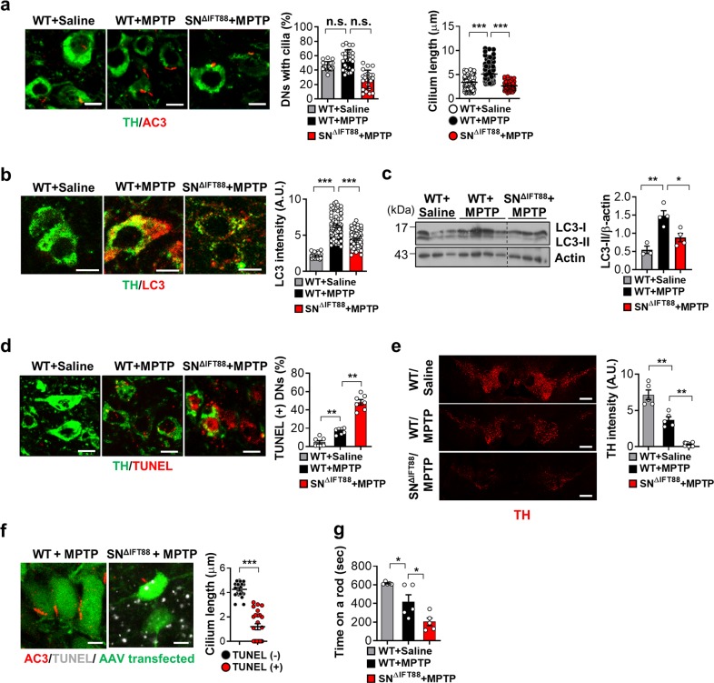
Fig. 7. Enhanced ciliogenesis in the substantia nigra dopamine neurons in the mice model of MPTP-induced Parkinson’s disease.
a Ciliary elongation in substantia nigra (SN) dopamine neurons (DNs) in the mouse after a single intraperitoneal administration of MPTP (30 mg/kg body weight) and blockade of cilia elongation with SN IFT88 knockdown. Two weeks before MPTP injection, the WT mice were injected with GFP-AAV and SN∆IFT88 mice were injected with IFT88-shRNA GFP-AAV into the bilateral SN. Primary cilia and DNs were stained using AC3 antibody and TH antibody. More than 100 TH-positive cells were analyzed in each animal. Scale bar, 10 μm. b, c Increased autophagy in SN DNs following MPTP treatment and blunted MPTP-induced autophagy in DNs with impaired ciliogenesis. Autophagy in DNs was evaluated by double staining with LC3 and TH antibodies and by LC3 immunoblotting. Scale bar, 10 μm. d Impaired ciliogenesis in the DNs enhances MPTP-induced apoptosis, as assessed by TUNEL staining. Scale bar, 10 μm. e MPTP treatment reduces the intensity of TH immunoreactivity in the SN pars compacta and this reduction is far greater in SNΔIFT88 mice. Scale bar, 500 μm. f Cilia elongation occurs before DN death. TUNEL-negative neurons of WT mice have long cilia but TUNEL-positive neurons in SNΔIFT88 mice have shorter or no cilia following MPTP treatment. Scale bar, 10 μm. g Motor function assessment using the rotarod test for 3 days after MPTP treatment in WT and SNΔIFT88 mice (n = 3~6 per group). Data are presented as the mean ± SEM. *p < 0.05, **p < 0.01, ***p < 0.005 between the indicated groups determined by ANOVA followed by a post hoc LSD test (n.s. = not significant).

