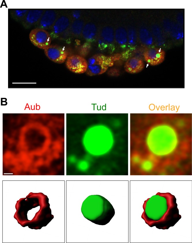
Figure 2.

Germ cells in cellular blastoderm embryos show large cytoplasmic granules with Aubergine shell around Tudor core. (A) An optical section of primordial germ cells formed at the embryo’s posterior co-stained with antibodies against Aub (red channel) and GFP-Tud (green channel). DAPI stains the nuclei of germ cells and somatic cells at the cortex of the embryo (blue channel). Large granules assembled in the cytoplasm of germ cells are indicated with arrows. (B) Top panels are optical sections obtained with super-resolution confocal microscopy which show Aub shell (red channel) wrapping around Tud core (green channel). Bottom panels are tilted 3D reconstructions of Aub shell (red), Tud core (green) and the composite granule internal segments to show the internal shell-core architecture of the granule imaged at the top panels. 10 internal optical sections were used for 3D reconstructions. Scale bar in (A) is 10 µm and in (B) it is 0.5 µm (see also Supplementary Fig. S2).
