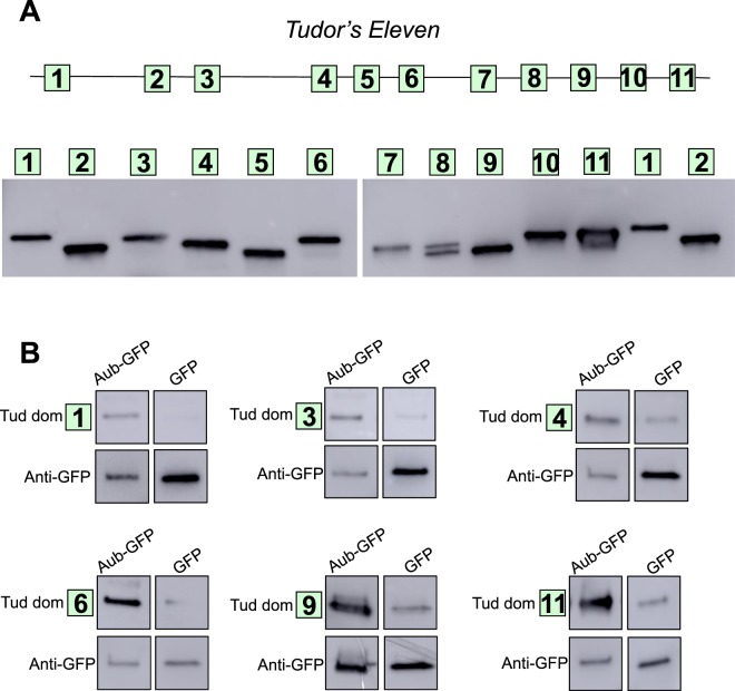
Figure 5.
Aubergine interacts with multiple Tudor domains distributed along the entire Tudor primary structure. (A) Top: a diagram of Tud protein with the location of all 11 Tud domains indicated as squares. Bottom: each Tud domain was individually expressed and partially purified for binding experiments with Aub. All Tud domains had a His tag at the N-terminus and were detected with anti-His tag antibody in a western-blot experiment. Right panel shows Tud domains 7–11 and domains 1 and 2, also shown on the left panel, for direct comparison of all Tud domains used for Aub binding experiments since two gels were used to accommodate all the domains. (B) Tud domains 1, 3, 4, 6, 9 and 11 bind to Aub in vitro. All Tud domains were added to anti-GFP beads associated with functional GFP-Aub purified from fly ovaries24,25 and bound domains were detected by western blot with anti-His tag antibody. Maternally-expressed GFP, purified from the ovaries in parallel with GFP-Aub, and bound to the anti-GFP beads was used as a negative control for the binding experiments. GFP proteins were detected with anti-GFP antibody. Each binding experiment is demonstrated as a group of four western-blot segments from the same gel showing a given Tud domain or GFP-Aub or just GFP. Corresponding full-length blots are shown in Supplementary Figs. S3 and S4. Full-length blots corresponding to binding experiments for all the other Tud domains that failed to bind to Aub are shown in Supplementary Figs. S5 and S6.

