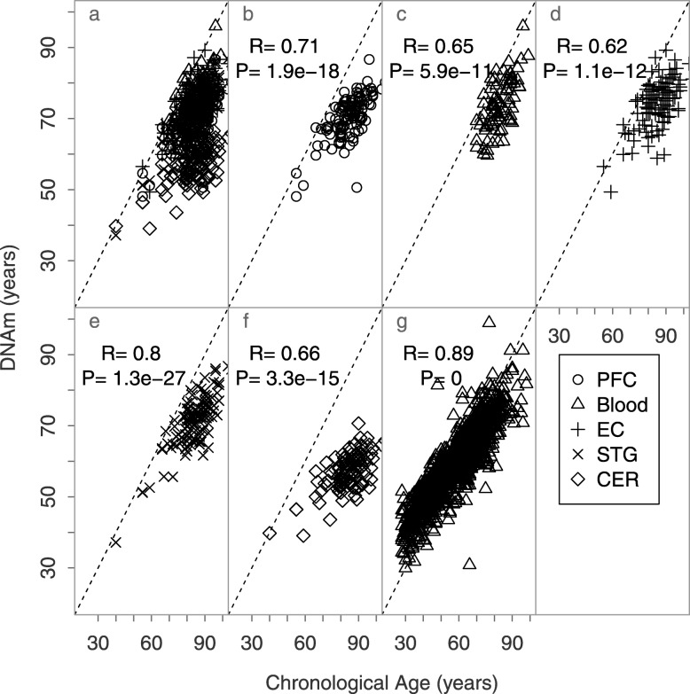
Fig. 1.
Scatterplots of chronological vs DNAm ages of brain and blood samples. Each point corresponds to an independent sample. The dotted line is the y = x bisector line, and the solid lines correspond to the regression line of each tissue. PFC, prefrontal cortex; EC, entorhinal cortex; STG, the superior temporal gyrus; CER, cerebellum (data from [9] for panels a–f and [10] for panel g)

