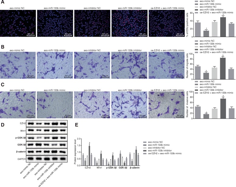
Fig. 6.
Exosomal miR-133b derived from MSCs represses glioma cell proliferation, migration, and invasion via inhibition of the Wnt/β-catenin signaling pathway. a Proliferation of glioma cells treated with exo-miR-133b mimic, exo-miR-133b inhibitor, or oe-EZH2 + exo-miR-133b mimic detected using EdU assay (200×). b Migration of glioma cells treated with exo-miR-133b mimic, exo-miR-133b inhibitor, or oe-EZH2 + exo-miR-133b mimic detected using Transwell assay (200×). c Invasion of glioma cells treated with exo-miR-133b mimic, exo-miR-133b inhibitor, or oe-EZH2 + exo-miR-133b mimic detected using Transwell assay (200×). d Protein band patterns of EZH2, Wnt1, p-GSK-3β, GSK-3β, and β-catenin detected in glioma cells treated with exo-miR-133b mimic, exo-miR-133b inhibitor, or oe-EZH2 + exo-miR-133b mimic. e Protein levels of EZH2, Wnt1, p-GSK-3β, GSK-3β, and β-catenin measured in glioma cells treated with exo-miR-133b mimic, exo-miR-133b inhibitor, or oe-EZH2 + exo-miR-133b mimic using Western blot analysis; #p < 0.05 vs. glioma cells treated with exo-mimic NC; &p < 0.05 vs. glioma cells treated with exo-inhibitor NC; measurement data were shown as mean ± standard deviation; comparisons among multiple groups were assessed by one-way analysis of variance (Dunnett’s post hoc test). The experiment was repeated three times to obtain the mean value

