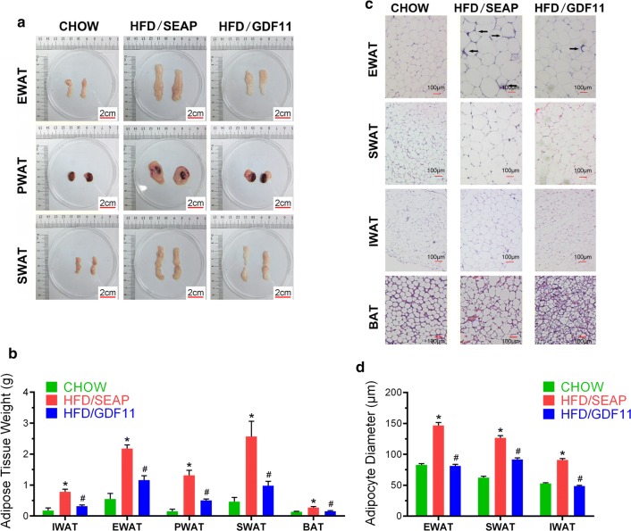
Fig. 2.
Impacts of Gdf11 gene transfer on adipose tissues. Adipose tissues were collected at the end of 8 weeks from animals fed regular Chow or HFD with hydrodynamic injection of pLIVE-GDF11 (HFD/GDF11) or control plasmids (HFD/SEAP). a Representative images of adipose tissues; b weight of fat pads; c histological images of EWAT, SWAT, IWAT and BAT (×10); d average diameter of adipocytes. SWAT subcutaneous WAT; IWAT inguinal WAT; PWAT perirenal WAT; EWAT epididymal WAT. Data represent mean ± SEM (n = 5). Arrows point to crown-like structures and scale bars represent 2 cm in a and 100 µm in c. *P < 0.05 comparing to mice fed regular Chow, #P < 0.05 comparing to HFD-fed control mice

