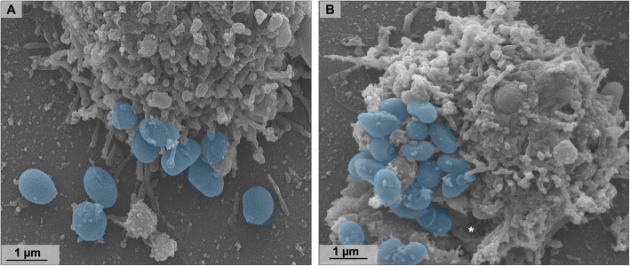
Fig. 10.
Orpheovirus particles are released from the cell both by exocytosis and lysis. a SEM at 24 h.p.i showing particles released from the cell by exocytosis (highlighted in blue). b SEM at 24 h.p.i showing particles released from the cell by lysis (highlighted in blue). Asterisk shows damage on cell surface, where particles seem to be released

