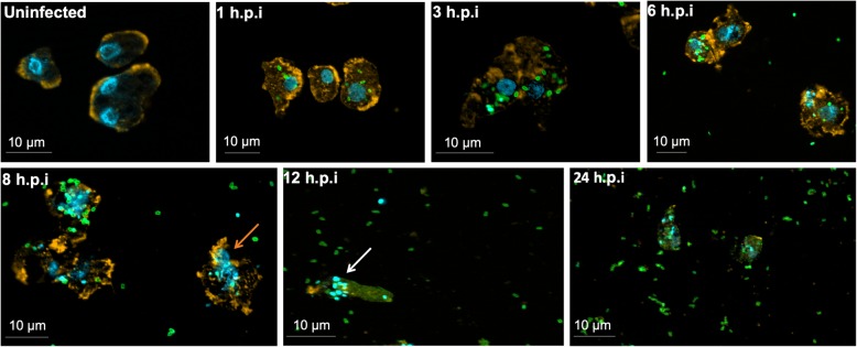
Fig. 2.
Characterization of the Orpheovirus replication cycle in V. vermiformis by IF. V. vermiformis monolayer was infected by Orpheovirus at an M.O.I. of 5 and visualized by IF. At early time points, Orpheovirus particles are observed attaching to the surfaces of amoebae. At 3 h.p.i. and 6 h.p.i., respectively, an increase in particle amounts can be visualized within the host cells. At 12 h.p.i., we noticed viral particle polarization at one cell extremity and an increase of viral particles outside the cells. At 24 h.p.i., many cells were rounded, and the large majority of amoebae were already lysed. The viral particles are in green (anti-orpheovirus particle antibody), amoeba cytoskeleton in orange (stained by rodamine-phalloidine) and the nucleus in blue (stained by DAPI). Scale bar, 10 μm

