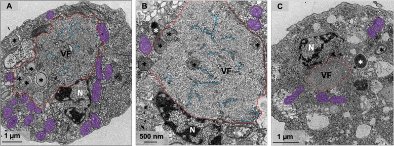
Fig. 4.
Electron-lucent viral factory and treatment with membrane trafficking inhibitor. a-b TEM image at 24 h.p.i shows that Orpheovirus present an electron-lucent VF (contoured in red and in detail), which occupy a large part of the host and is observed in perinuclear regions. We also observed membrane recruitment inside the VF (blue), and the host nucleus remains present during the infection. c Treatment with BFA impacts both the formation of VFs and morphogenesis of new particles. VF: viral factory; N: nucleus; Mitochondria are highlighted in purple; *viral particles

