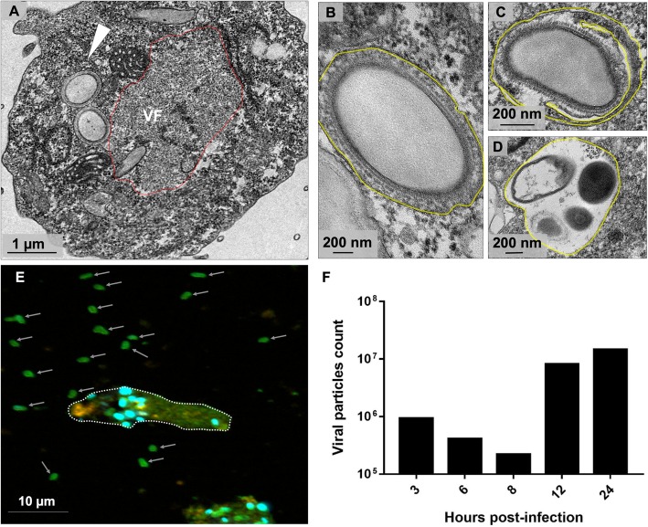
Fig. 9.
Orpheovirus particles are released from the cell by exocytosis. a TEM image at 24 h.p.i evidencing new particles in peripheral regions in the host cell cytoplasm (white arrow). b and c Some particles were observed involved by one or more membranes (highlighted in yellow). d More than one particle in the same vacuole were evidenced. e IF assays at 12 h.p.i showing fusiform cells and an increase of particles outside the host cells released by exocytosis (white arrows). f Count of Orpheovirus particles in the supernatant over the replication cycle (M.O.I. = 5)

