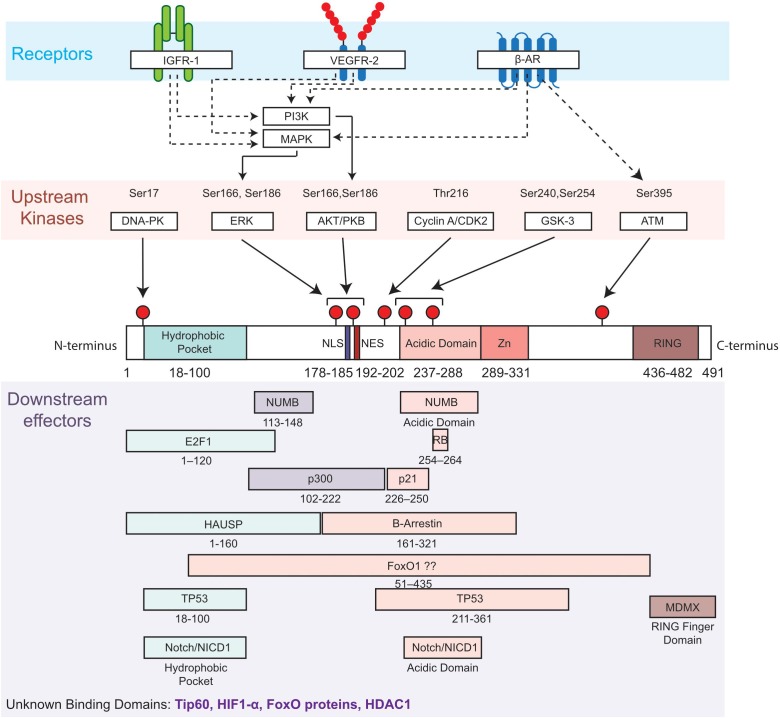
FIGURE 1.
Diagram of MDM2 domains showing phosphorylation sites and upstream regulators (kinases and related receptors) of MDM2 functions, as well as putative binding sites with downstream effectors. Top, most common phosphorylation sites on Serine (Ser) and Threonine (Thr) are indicated by a red pin. Residues are numbered based on amino acid sequence from N-terminus to C-terminus. Middle, the functional domains of MDM2 include the hydrophobic pocket, the nuclear localization sequence (NLS), the nuclear export sequence (NES), the acidic and central domain, the Zinc domain (Zn) and the ring domain (RING). Bottom, selected interactions between MDM2 and its downstream effectors of MDM2 are illustrated. β-Adrenergic receptor, β-AR; DNA-dependent protein kinase, DNA-PK; E2F transcription factor 1, E2F1; extracellular-signal-regulated kinase, ERK; Forkhead box protein, FoxO; herpes virus-associated ubiquitin specific protease, HAUSP; Insulin-like growth factor receptor 1, IGFR1; Mitogen-activated protein kinases, MAPK; Mouse double Minute 4, MDM4; NUMB; Notch intracellular domain, NICD; Phosphotidylinositol 3-kinase, PI3K; Protein kinase B, PKB; protein numb homolog, Retinoblastoma protein, RB; Vascular Endothelial Growth Factor Receptor 2, VEGFR2.

