
Figure 5.
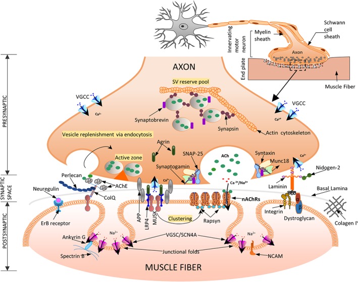
A schematic representation of a neuromuscular junction (NMJ) and its main molecular actors—three specific regions define the NMJ: (a) the presynaptic motor nerve terminal where vesicles fuse with the terminal membrane to release acetylcholine (ACh) into the synaptic cleft. Calcium influx through the voltage‐gated Ca channels (VGCC) trigger vesicle fusion and release from the active zones (described in detail in Section 5.1); (b) the synaptic space contains the basal lamina (BL, extra cellular matrix layer), and shows the presence of AChE‐ColQ (essential for the inactivation of ACh). ColQ binds MuSK and Perlecan necessary for stabilization of BL. MuSK enables AChR clustering via rapsyn (detailed in Section 5.2); (c) postsynaptic organization of the skeletal muscle membrane include several folds with receptors for the diffusing ACh (AChRs) at the crest and voltage‐gated sodium channels (VGSC) in the troughs of the folds necessary for efficient neuromuscular transmission. The agrin‐Lrp4‐MuSK complex, present on the trough of the postsynaptic membrane is essential for the formation of the NMJ (described in detail in Section 5.3). The entire structure is finally attached to the actin cytoskeleton (not shown here for simplicity)