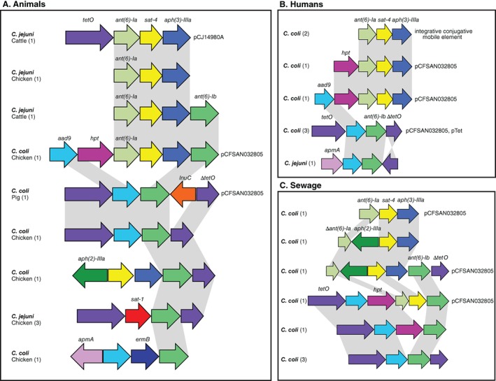
Figure 3.

Comparative genetic organization of AMR GIs in Campyloabcter. The presence of each AMR gene, highlighted in different colours, is shown for representative C. jejuni and C. coli isolate genomes sampled from animals (A), humans (B) and sewage (C). The number of isolate genomes containing each genomic island arrangement is indicated in the parenthesis. Grey shading identifies sequence that shares > 95% nucleotide sequence identity. The name of the plasmid or mobile genetic element, associated with each genomic island, is indicated. [Color figure can be viewed at http://wileyonlinelibrary.com]
