Figure 2.

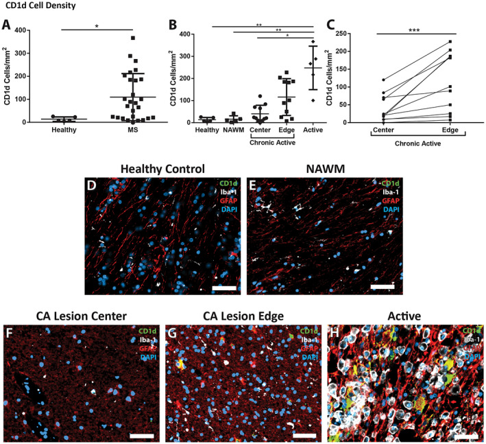
Changes in the density of CD1d in MS patients. A. Multiple sclerosis tissue showed a significant increase in density of CD1d‐positive cells when compared to controls (n = 27, and 5, respectively). B. The increase in CD1d‐postive cell density was significantly different in different lesion types, and different areas within those lesions. Active lesions had a much greater density than chronic active lesion centers, NAWM, and controls (n = 5, 11, 5 and 5, respectively). The edges of chronic active lesions had a higher density of CD1d‐positive cells than controls and NAWM, though did not achieve significance (n = 11, 5 and 5, respectively). C. Density of CD1d was significantly greater in the chronic active lesion edge than in the paired chronic active lesion center (***P = 0.001, n = 11 per group, Wilcoxon matched‐pairs signed‐rank test). Immunofluorescence shows highly reactive astrocytes (red, GFAP), but not microglia (white, Iba‐1), immunoreactive for large amounts of CD1d (green) in the active lesion (H) with double‐labeling appearing as yellow, while no CD1d is evident in the control (D) or NAWM (E). The chronic active lesion center (F) and chronic active lesion edge (G) demonstrate a more intermediate astrocyte phenotype (less hyperplastic and more fibrillary), though there are appreciable levels of CD1d in the reactive astrocytes within the chronic active lesion edge. DAPI (blue) as nuclear stain; scale bars = 50 µm. *P ≤ 0.05, **P ≤ 0.01. Bars represent the mean, and error bars the standard deviation. (A, C) Mann‐Whitney, (B) Kruskal‐Wallis with Dunn's multiple comparison test.
