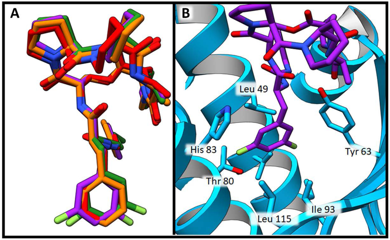
Figure 6.
Depsipeptide phenylalanine motif binding pocket. A) Structure overlay demonstrating similarity of phenylalanine binding positions. Compound 2 is shown in red, compound 5 is shown in orange, compound 16 is shown in purple and ADEP4 is shown in green. B) Binding pocket of compound 16 shown in purple. SaClpP is shown in blue ribbon structure. SaClpP side chains within 4 Å of the phenyl ring of this ligand are shown as blue sticks.

